
New
-3%
glimz
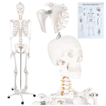
Glimz human skeleton anatomical model 170 cm with stand
Delivery in 5–14 business days
160.90 €165.70 €
Anatomical models include glimz and 4Fizjo human skeletons alongside dental, jaw, spine, eye, ear and ankle joint models. They suit anatomy study and visual explanations during lessons or consultations. Choose according to the body part being discussed, the scale required and the features to demonstrate, such as removable teeth, implants, braces or joint ligaments.
Human anatomical models cover the skeleton, spine, teeth and jaw, as well as the eye, ear and ankle joint. The range includes glimz and 4Fizjo models for different teaching and demonstration topics. A complete skeleton provides an overview of bone arrangement, while a model of an individual body part keeps attention on a specific anatomical area.
Start with the subject of the explanation: overall skeletal structure, ligament placement, dental conditions or orthodontic braces. Then compare the stated dimensions, scale and demonstration features. A skeleton needs more display space than a dental jaw model. An enlarged organ model supports discussion of its structure, while a life-size model helps relate the represented body part to human proportions.
For lessons, anatomy revision or consultations, match the model to the topic being presented. Dental models support explanations involving teeth and jaws, while skeleton, spine and ankle models serve different aspects of skeletal anatomy.
The dental model should match the topic being explained. Options with implants, braces, dental pathologies or removable teeth support different dental demonstrations.
A 1:1 anatomical model represents the body part at life size. The spine and ankle joint models include options described with this scale.
Consider the skeleton model's stated height and whether a stand is included. Allow space for the model and for people to view it during a demonstration.
+371 20408041
info@motivs.lv
Duntes 56, Rīga, LV-1005, Latvija
Monday: 10:00 - 19:00
Tuesday: 10:00 - 19:00
Wednesday: 10:00 - 19:00
Thursday: 10:00 - 19:00
Friday: 10:00 - 19:00
Saturday: Closed
Sunday: Closed
 Gudri pirkumi vienuviet